
Date : 11/18/2026
Disclaimer: This article is for general educational purposes only and isn’t a substitute for professional medical advice, diagnosis or treatment. Only your physician and radiology team can decide whether gadolinium contrast is right for you. Before your scan, tell them if you have kidney disease, have had a past reaction to contrast, or are pregnant or breastfeeding. If you have a medical emergency, call your local emergency number right away.
Table of Contents
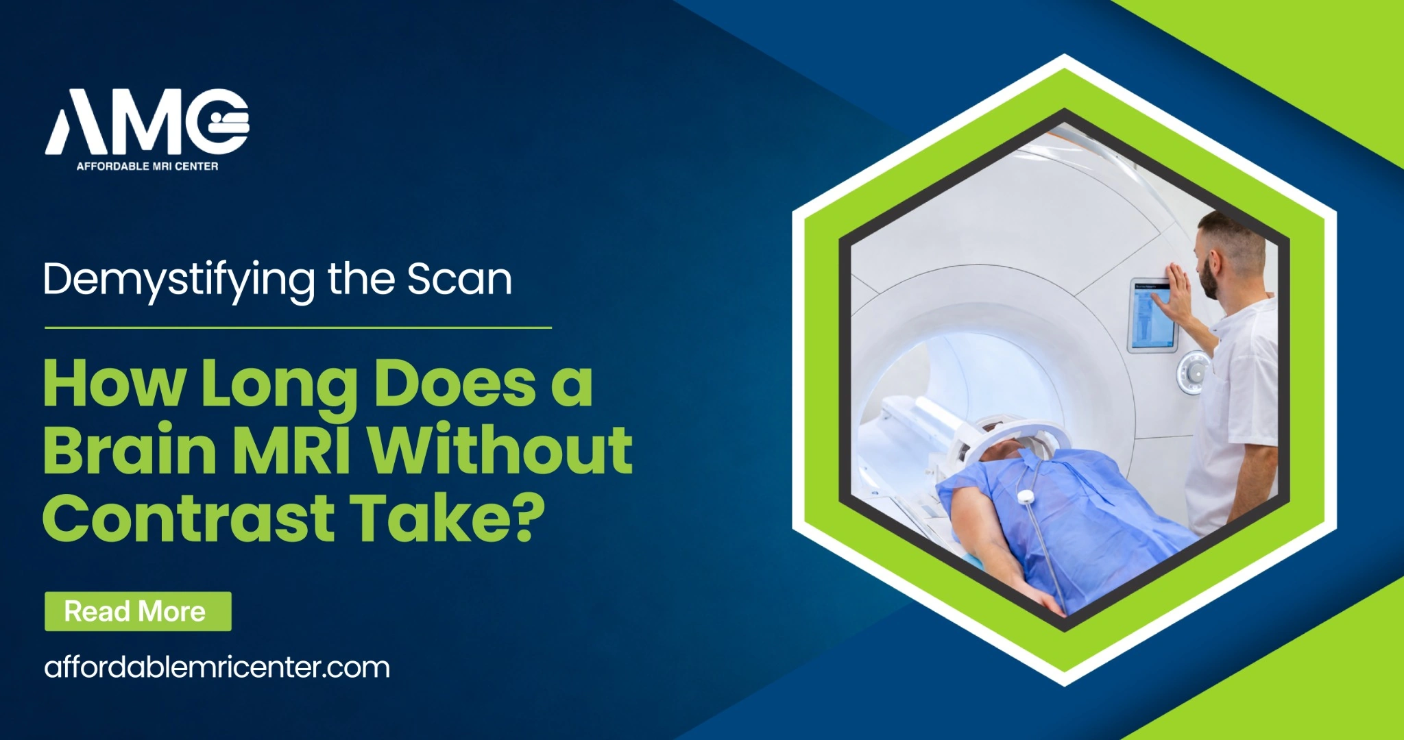
Facing a neurological scan is inherently nerve-wracking. The sterile room. That giant, humming cylindrical machine. It naturally spikes anxiety. Before sliding into that narrow tube, one specific question dominates almost every patient’s mind: how long does a brain mri without contrast take? You want a precise timeline. Because knowing exactly how many minutes you will spend holding perfectly still is the ultimate defense against claustrophobia and pre-appointment panic.
Book Your Appointment Now
The Minute-by-Minute Reality: How Long Does a Brain MRI Without Contrast Take?
Let’s cut straight to the chase. If you are stripping away the preparation, the paperwork, and the post-scan dressing time, the actual time spent actively inside the magnetic bore is remarkably brief. For a standard, uncomplicated neurological scan without any dye, the machine captures images for roughly 15 to 30 minutes.
That said, total appointment times look a bit different. When patients ask how long does a brain MRI without contrast take, they often confuse the active scanning duration with the total facility visit. You should absolutely plan to be at the imaging center for about 45 minutes to an hour.
Why the discrepancy? The timeline involves several critical safety and positioning steps that must happen before the magnetic coils ever power up. Technologists have to conduct a rigorous safety screening. You cannot bring any ferromagnetic metals near a 1.5T or 3.0T magnet. That means stripping off jewelry, hairpins, watches, and even certain athletic wear that contains hidden metallic anti-microbial threads.
Once cleared, you are positioned on the sliding table. The technologist will place a specialized device called a head coil over your face. Think of it like a highly sensitive antenna helmet; it captures the radio frequency signals bouncing off your tissues. After securing ear protection—because the machine gets incredibly loud—you are slid into the isocenter of the magnet.
The Standard Timeline Breakdown
To paint a perfectly clear picture of your upcoming appointment, here is how those minutes actually break down in a clinical setting.
| Appointment Phase | Estimated Duration | What Actually Happens |
|---|---|---|
| Safety & Screening | 10 – 15 Minutes | Medical history review, safety questionnaire, changing into metal-free facility scrubs. |
| Physical Positioning | 5 Minutes | Lying down, securing earplugs or headphones, placing the head coil, and getting the emergency panic button. |
| Active Scanning | 15 – 30 Minutes | The actual imaging sequence. Expect loud knocking, heavy buzzing, and rhythmic thumping sounds. |
| Post-Scan Discharge | 5 Minutes | Sliding out of the tube, changing back into street clothes, and heading home. |
Decoding the Jargon: How Long Does a Brain MRI without Contrast Take?
Medical terminology often feels designed to confuse patients. When you glance at your physician’s script and spot the abbreviation “w/o contrast,” it simply translates to “without contrast.”
But to really grasp the w/o contrast meaning, you have to understand what is physically being left out of your procedure. Contrast refers to a gadolinium-based dye. It is a heavy metal compound injected intravenously (directly into an arm vein) midway through a scan. This chemical alters the magnetic properties of nearby water molecules inside your body, which effectively brightens blood vessels or lesions on the final black-and-white images.
A “without contrast” study skips the needle entirely. No IV. No metallic taste in your mouth. The imaging technology relies 100% on the natural tissue properties of your brain—using specific radio wave sequences (like T1 and T2 weighting) to clearly differentiate between gray matter, white matter, and cerebrospinal fluid.
Diagnostic Accuracy: Is MRI Without Contrast Effective?
Anxiety often creeps in when patients realize they are skipping the dye. A frequent concern is that skipping the injection means getting a lower-quality evaluation. People understandably ask: Is MRI without contrast effective?
The medical consensus is a definitive yes.
For the vast majority of routine neurological complaints, a non-contrast scan is the ultimate gold standard baseline test. Modern MRI machines possess astonishing spatial resolution. They detect a massive array of severe, life-altering conditions without needing a single drop of chemical enhancement.
Neurologists rely on standard non-contrast sequences to instantly evaluate:
- Acute Ischemic Strokes: Using a highly sensitive technique called diffusion-weighted imaging (DWI), a standard MRI spots a stroke within minutes of symptom onset.
- Traumatic Brain Injuries: It easily highlights concussion damage, deep tissue contusions, or minor hematomas following a nasty fall or car accident.
- Structural Anomalies: Things like congenital defects, hydrocephalus (fluid buildup), or brain shrinkage (atrophy) linked to early-stage dementia.
- Multiple Sclerosis (Initial Diagnosis): While doctors use contrast later to check for active flare-ups, the baseline scars (demyelinating lesions) are entirely visible without any dye.
So, if a patient presents with chronic migraines, unexplained dizziness, or baseline memory loss, skipping the IV does absolutely nothing to compromise the diagnostic integrity of the scan.
Book your Appointment
Digging Deeper: What Will an MRI With Contrast Show?
After you have figured out how long does a brain MRI without contrast take, its time to know exactly what it is capable of. Even with the immense power of a standard scan, certain clinical mysteries require a heavier diagnostic hammer. Standard tissue imaging eventually hits a ceiling. So, what will an MRI with contrast show that a regular one simply cannot?
Gadolinium acts like a neon highlighter for the vascular system and the blood-brain barrier. In a perfectly healthy brain, this barrier acts as a microscopic security gate. It tightly restricts what enters the brain tissue from the bloodstream, keeping the gadolinium dye safely locked inside the blood vessels.
However, catastrophic diseases break that barrier down. Aggressive brain tumors, severe viral infections (like encephalitis), and active demyelinating diseases compromise this cellular wall. When the blood-brain barrier breaks, the injected dye leaks out into the surrounding brain tissue. On the radiologist’s monitor, that leaked dye lights up intensely white. This precise lighting allows surgeons to map the exact, jagged boundaries of a tumor, see the blood vessels feeding it, or confirm that an MS lesion is actively inflamed today, rather than an old scar from years ago.
Clinical Reasoning: Why Would a Doctor Order an MRI With Contrast?
Because a contrast scan requires IV insertion, nearly doubles the time in the machine, and carries minor risks (such as allergic reactions or kidney processing issues), physicians never order it casually.
Understanding exactly why would a doctor order an MRI with contrast comes down to the absolute necessity for granular, microscopic detail regarding cellular activity and blood flow. A physician will specifically write a script for contrast if they are hunting for:
- Cancerous Growths: To differentiate between a harmless, benign cyst and a malignant tumor, and to plan precise surgical margins.
- Deep Infections: To pinpoint dangerous brain abscesses or severe inflammation of the meninges (meningitis).
- Post-Operative Checks: To figure out if a suspicious spot on the scan is just normal, healing surgical scar tissue, or a recurring tumor coming back.
- Complex Vascular Issues: Using a technique called an MRA (Magnetic Resonance Angiography) with contrast to map out the intricate web of brain arteries and spot weakened, ballooning blood vessels (aneurysms) before they rupture.
The Clinical Comparison: With vs. Without
| Feature | The Non-Contrast Scan | The Contrast-Enhanced Scan |
|---|---|---|
| Primary Clinical Use | Headaches, dizziness, trauma, stroke, and basic structural mapping. | Tumors, brain infections, active MS flare-ups, and complex vascular mapping. |
| Active Scanner Time | 15 – 30 Minutes. | 45 – 60 Minutes (Requires two completely separate imaging runs). |
| Invasive Elements | Completely non-invasive. | Requires an IV line and gadolinium injection. |
| Preparation Needs | Remove all metals, wear scrubs. | May require strict fasting or a prior blood test (GFR) to verify healthy kidney function. |
Motion Artifacts: Does Fidgeting Change How Long Does a Brain MRI Without Contrast Take?
The short answer? Yes, absolutely.
When mapping out how long does a brain MRI without contrast take, that 15-to-30-minute window comes with a massive caveat: the patient must remain entirely frozen. The machine captures images in “slices.” If you swallow hard, cough, twitch your nose, or wiggle your toes, those micro-movements ripple up to your head.
The result? The image blurs. Radiologists call these “motion artifacts.” When an image blurs, it is diagnostically useless. The technologist operating the machine from the other room has no choice but to delete that specific sequence and start it over. A sequence can last anywhere from two to six minutes. Fidgeting just three times can easily tack an extra 15 minutes onto your scan.
To get out of the tube as fast as possible, close your eyes before you even enter the machine. Keep them shut. Focus entirely on slow, rhythmic belly breathing.
Conclusion
Frequently Asked Questions
Claustrophobia itself does not alter the machine’s speed, but panic attacks will halt the scan entirely. If a patient presses the emergency squeeze ball, the magnetic sequences stop, and the patient is pulled out. To prevent this, individuals with severe claustrophobia are routinely prescribed a mild oral sedative (like Valium or Xanax) by their referring doctor to take an hour before the appointment. Provided the patient remains calm and still, the scan still only takes 15 to 30 minutes.
If your medical order explicitly says “without contrast,” fasting is rarely required. You can usually eat breakfast, drink water, and take your standard daily medications. Fasting protocols are generally reserved for abdominal imaging or contrast-enhanced scans to prevent nausea when the dye hits the bloodstream.
Typically, imaging centers will say no. Even if your favorite leggings or sweater feels soft and lacks zippers, modern athletic and lounge wear heavily utilizes micro-threads of silver or copper to kill sweat bacteria. Those invisible metals heat up rapidly under magnetic radio frequencies, causing genuine skin burns. Slipping into provided facility scrubs is the only way to guarantee 100% safety.
No, your head is never mechanically locked, bolted, or strapped tightly to the table. However, it will be situated inside the head coil—which feels a bit like a loose-fitting fencing mask. Technologists will often wedge soft acoustic foam sponges between your ears and the hard plastic of the coil. This serves a dual purpose: it heavily muffles the deafening clanging of the machine, and it acts as a gentle physical reminder to keep your skull perfectly still.
References
https://my.clevelandclinic.org/health/diseases/24208-ischemic-stroke-clots
https://pmc.ncbi.nlm.nih.gov/articles/PMC9047866/
Dr. Samiullah Kundi
Neurology, Pain Medicine & Neuroimaging
Dr. Kundi is a board-certified neurologist and pain medicine specialist who relies on advanced MRI findings to diagnose and treat complex conditions of the brain, spine and peripheral nerves. He is certified by the American Board of Psychiatry and Neurology (ABPN) in Neurology and Clinical Neurophysiology, the American Board of Pain Medicine (ABPM) and the American Board of Integrative Holistic Medicine (ABIHM). He believes precise, timely imaging is the foundation of effective care, and that belief led him to support Affordable MRI Center’s mission of making high-quality diagnostic MRI accessible and affordable.