
Date : 07/08/2026
Disclaimer : The information provided in this article is for educational and informational purposes only and is not intended as a substitute for professional medical advice, diagnosis, or treatment. This guide is not a substitute for a physical examination or a formal review of your MRI images by a licensed medical doctor. Never disregard professional medical advice or delay in seeking it because of something you have read here. If you think you may have a medical emergency, call your doctor or emergency services immediately.
Table of Contents
Dealing with sharp, shooting leg pain is exhausting. Simple things like sitting at a desk, driving to work, or even just trying to sleep become unbearable. People want answers, and they want them fast. The big question on most minds is this: can MRI diagnose sciatica accurately? Yes. Magnetic Resonance Imaging is a phenomenal tool. It lets doctors look right past the bone to see exactly where a nerve is getting crushed.
This guide dives deep into nerve pain anatomy. It covers the clinical steps taken before imaging. It also breaks down exactly what doctors look for on the screen when trying to put an end to your pain.
The Reality of Sciatica and Nerve Anatomy
Sciatica isn’t actually a disease. It’s a symptom. It points to a mechanical failure happening higher up in the lower back. The sciatic nerve is huge. It is, in fact, the longest and thickest nerve in the human body. Five distinct nerve roots in the lumbar spine and sacrum (specifically L4, L5, S1, S2, and S3) merge together. They form one massive nerve bundle. This bundle travels deep through the gluteal muscles. Then, it runs all the way down the back of both legs, branching out into the calves and feet.
When something pinches this nerve pathway, things go wrong quickly. Symptoms hit hard.
- Intense, electric-shock pain firing down one leg.
- Numbness. That awful “pins and needles” feeling in the foot or calf that just won’t fade.
- Leg weakness. You might notice your leg giving out unexpectedly while walking up stairs.
- Pain that spikes aggressively when you cough, sneeze, or sit in one position for too long.
Because the nerve is so incredibly long, a tiny pinch at the top translates to massive pain at the bottom. A localized problem deep inside the spine makes your toes go numb. It is a frustrating reality for patients. Understanding this anatomy is crucial because it explains why treating the leg itself rarely works. The fire is in the leg, but the spark is in the spine.
From Symptoms to Scans: The Initial Steps
Before ordering expensive imaging, doctors need context. When wondering if can MRI diagnose sciatica in a specific patient, clinicians first need to map the pain. The journey starts in the exam room. Not the radiology tube.
Clinical Assessments and Physical Tests
A thorough physical examination for sciatica is always the first move. Doctors use specific body movements to figure out which nerve root is screaming. They look at your posture, how you walk, and where exactly the pain travels.
The Straight Leg Raise (SLR) is the classic diagnostic method. A patient lies completely flat on their back. The doctor slowly lifts the painful leg upwards. If shooting pain happens between 30 and 70 degrees of elevation? That is a massive red flag. It usually points straight to a herniated disc putting pressure on the lower lumbar nerves.
But that isn’t the only check. Other sciatica diagnosis tests happen right there on the exam table. Reflexes get checked with a small rubber hammer. A sluggish knee or ankle jerk reflex tells a story. Muscle strength is tested. Patients might be asked to walk on their heels or tiptoes. If you can’t walk on your toes, the S1 nerve root is likely compressed. Sensory checks find the exact dead spots of numbness on the skin.
These physical tests for sciatica are highly accurate at proving nerve involvement. Once the doctor knows the nerve is genuinely angry, they decide if looking closer with advanced technology is necessary.
How Can MRI Diagnose Sciatica?
Physical exams prove a pinch exists. Imaging shows the exact location and the culprit. This is where advanced radiology truly shines.
An MRI machine uses incredibly powerful magnets and radio waves. It builds detailed cross-sections of the body. Standard X-rays only show bone. An MRI shows the soft stuff. Spinal cords, individual nerves, delicate discs, tough ligaments. It reveals it all. That makes it undeniably the best scan for sciatica.
Patients constantly ask, does sciatica show up on MRI? Well, pain itself doesn’t glow on a computer screen. But the structural disaster causing the pain absolutely does.
What the Doctor Actually Sees
Looking at a lumbar scan, specialists hunt for specific things. They analyze the black, white, and gray images for abnormalities.
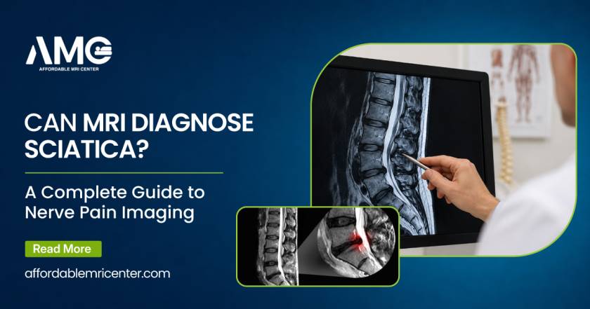
- Disc Herniations: The classic culprit. The jelly-like center of a spinal disc bursts through its tough outer ring. The scan shows exactly how hard that jelly is smashing into the adjacent nerve root.
- Spinal Stenosis: The bony canal holding the nerves gets too narrow. Usually, bone spurs or thick ligaments are to blame. The MRI shows the spinal cord getting choked in that tight space.
- Spondylolisthesis: One spinal bone slips forward over another. It acts like a guillotine on the nerve.
- Cysts and Tumors: Rare. But when they happen, an MRI catches them instantly.
So, can MRI diagnose sciatica early on? Yes, but doctors usually wait. Why? Because lots of people have ugly-looking discs on an MRI but zero pain. Bodies age. Discs bulge naturally over time. The images must match the physical symptoms perfectly. If a scan shows a bulging disc on the left side, but your pain is on the right, that disc isn’t the problem.
Comparing Your Imaging Options
It is incredibly common to wonder about getting an xray for sciatica. Patients want quick answers. X-rays are fast. They are cheap. You can get one at almost any urgent care clinic. But they are nearly useless for nerve pain. They simply cannot see soft tissue.
Here is a breakdown of how the different imaging tools stack up.
Imaging Modalities for Nerve Pain
| Tool | What It Does Best | The Downside for Nerve Pain | When It Is Used |
|---|---|---|---|
| X-Ray | Viewing bones, arthritis, severe fractures. | Cannot see discs or nerves at all. Misses nerve compression entirely. | Ruling out broken bones or massive spinal instability. |
| CT Scan | Highly detailed 3D bone structures. | Terrible soft tissue detail. Exposes the patient to radiation. | Used if an MRI is too dangerous (e.g., patient has a certain pacemaker). |
| MRI Scan | Crystal clear views of discs, nerves, and ligaments. | Almost too detailed. Might show normal aging issues that aren’t causing the actual pain. | Finding the exact millimeter where a nerve is being crushed. |
Finding the Root Cause of the Pain
If you are asking can you see sciatica on MRI, you are really asking if the doctor can see the underlying pathology. The answer is overwhelmingly yes. Advanced imaging takes the guesswork out of treatment planning.
This next table highlights the most common villains found on a lumbar scan.
Common Structural Causes of Nerve Compression
| The Condition | What Is Actually Happening | How It Looks on the Screen |
|---|---|---|
| Herniated Disc | Disc nucleus leaks outward. | A dark blob aggressively pushing into the bright spinal fluid and nerve. |
| Degenerative Disc Disease | Discs dry out and collapse with age. | Discs look dark (no water) and completely flattened. Nerves lose their exit space. |
| Lumbar Spinal Stenosis | The main nerve canal shrinks. | Thick ligaments or overgrown bone crowding out the spinal canal. |
| Piriformis Syndrome | A muscle in the buttock spasms. | Sometimes shows up as muscle swelling. Usually diagnosed by physical exam, not imaging. |
Given the sheer amount of detail provided by the scan, there is zero doubt that can MRI detect sciatica triggers with pinpoint precision. It is the ultimate roadmap for surgeons, physical therapists, and pain management specialists.
The Treatment Journey After the Scan
Once the scan results are in, the guessing games end. The medical team finally knows what they are up against. If a massive herniated disc is choking the nerve, physical therapy can be tailored specifically to pull that disc material away from the nerve root. If spinal stenosis is the issue, treatment might lean toward epidural steroid injections to flush the cramped area with heavy anti-inflammatory medication.
The beauty of a clear diagnosis is that it prevents wasted time. Nobody wants to spend months doing the wrong stretches for a misdiagnosed back issue. The imaging provides a factual baseline. If surgery becomes necessary down the line, the surgeon already has a precise map of where to operate.
Conclusion
Frequently Asked Questions
Yes. While the machine does not literally “see” your pain, it clearly highlights the physical problems causing the nerve compression. Whether it is a herniated disc, a bone spur, or a thickened ligament, the scan reveals the exact source of the irritation.
Usually, no. Most sudden nerve pain fixes itself within a month to six weeks. Physical therapy helps. Time helps. Anti-inflammatory medication helps. Scans are saved for pain that stubbornly refuses to leave. They are also ordered immediately if terrifying “red flags” pop up—like suddenly dropping objects, severe leg weakness, or losing control of your bladder.
The machine itself? Not at all. It uses strong magnets and radio waves, meaning there is zero radiation. It is completely painless. The hard part is lying still. When your leg is on fire with severe nerve pain, 30 to 45 minutes flat on a hard table feels like an absolute eternity. If you are in severe pain, doctors can sometimes prescribe a mild muscle relaxer or sedative beforehand to help you get through the scan comfortably.
It happens more than you might think. It can be incredibly frustrating to get a “normal” result when you are in agony. But it is actually helpful data. If the lower back looks perfect, the pinch is happening somewhere else. The sciatic nerve runs a very long way. It might be getting squeezed down in the glutes by a spasming muscle. Doctors use this “normal” result to pivot their strategy and look further down the leg.